Abdominal Aortic Aneurysm
What is an abdominal aortic aneurysm?
 |
| Click Image to Enlarge |
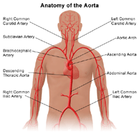
An abdominal aortic aneurysm, also called AAA, is a bulging, weakened area in the wall of the aorta (the largest artery in the body) resulting in an abnormal widening or ballooning greater than 50 percent of the normal diameter (width).
The aorta extends upward from the top of the left ventricle of the heart in the chest area (ascending thoracic aorta), then curves downward (aortic arch) through the chest area (descending thoracic aorta) into the abdomen (abdominal aorta). The aorta delivers oxygenated blood pumped from the heart to the rest of the body.
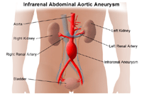
The most common location of arterial aneurysm formation is the abdominal aorta, specifically, the segment of the abdominal aorta below the kidneys. An abdominal aneurysm located below the kidneys is called an infrarenal aneurysm. An aneurysm can be characterized by its location, shape, and cause.
 |
| Click Image to Enlarge |
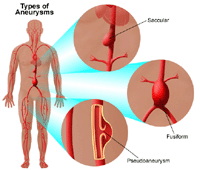
The shape of an aneurysm is described as being fusiform or saccular which helps to identify a true aneurysm. The more common fusiform shaped aneurysm bulges or balloons out on all sides of the aorta. A saccular shaped aneurysm bulges or balloons out only on one side.
A pseudoaneurysm, or false aneurysm, is an enlargement of only the outer layer of the blood vessel wall. A false aneurysm may be the result of a prior surgery or trauma. Sometimes, a tear can occur on the inside layer of the vessel resulting in blood filling in between the layers of the blood vessel wall creating a pseudoaneurysm.
The aorta is under constant pressure as blood is ejected from the heart. With each heart beat, the walls of the aorta distend (expand) and then recoil (spring back), exerting continual pressure or stress on the already weakened aneurysm wall. Therefore, there is a potential for rupture (bursting) or dissection (separation of the layers of the aortic wall) of the aorta, which may cause life-threatening hemorrhage (uncontrolled bleeding) and, potentially, death. The larger the aneurysm becomes, the greater the risk of rupture.
 |
| Click Image to Enlarge |
Because an aneurysm may continue to increase in size, along with progressive weakening of the artery wall, surgical intervention may be needed. Preventing rupture of an aneurysm is one of the goals of therapy.
What causes an abdominal aortic aneurysm to form?
An abdominal aortic aneurysm may be caused by multiple factors that result in the breaking down of the well-organized structural components (proteins) of the aortic wall that provide support and stabilize the wall. The exact cause is not fully known.
Atherosclerosis (a build-up of plaque, which is a deposit of fatty substances, cholesterol, cellular waste products, calcium, and fibrin in the inner lining of an artery) is thought to play an important role in aneurysmal disease, including the risk factors associated with atherosclerosis, such as:
-
age (greater than 60)
-
male (occurrence in males is four to five times greater than that of females)
-
family history (first degree relatives such as father or brother)
-
genetic factors
-
hyperlipidemia (elevated fats in the blood)
-
hypertension (high blood pressure)
-
smoking
-
diabetes
Other diseases that may cause an abdominal aneurysm include:
-
genetic disorders of connective tissue (abnormalities that can affect tissues such as bones, cartilage, heart, and blood vessels), such as Marfan syndrome, Ehlers-Danlos syndrome, Turner’s syndrome, and polycystic kidney disease
-
congenital (present at birth) syndromes, such as bicuspid aortic valve or coarctation of the aorta
-
giant cell arteritis - a disease that causes inflammation of the temporal arteries and other arteries in the head and neck, causing the arteries to narrow, reducing blood flow in the affected areas; may cause persistent headaches and vision loss
-
trauma
-
infectious aortitis (infections of the aorta) due to infections such as syphilis, salmonella, or staphylococcus. These infectious conditions are rare.
What are the symptoms of abdominal aortic aneurysms?
Abdominal aortic aneurysms may be asymptomatic (without symptoms) or symptomatic (with symptoms).
About three of every four abdominal aortic aneurysms are asymptomatic and may be found upon routine physical examination by the discovery of a pulsating mass in the abdomen. An aneurysm may also be discovered by x-ray, computed tomography scan (CT scan), or magnetic resonance imaging (MRI) that is being done for other conditions. Since abdominal aneurysm may be present without symptoms, it is referred to as the “silent killer” because it may rupture before being diagnosed.
Pain is the most common symptom of an abdominal aortic aneurysm. The pain associated with an abdominal aortic aneurysm may be located in the abdomen, chest, lower back, or groin area. The pain may be severe or dull. The occurrence of pain is often associated with the imminent (about to happen) rupture of the aneurysm.
Acute, sudden onset of severe pain in the back and/or abdomen may represent rupture and is a life threatening medical emergency.
The symptoms of an abdominal aortic aneurysm may resemble other medical conditions or problems. Always consult your physician for more information.
How are aneurysms diagnosed?
In addition to a complete medical history and physical examination, diagnostic procedures for an aneurysm may include any, or a combination, of the following:
-
computed tomography scan (Also called a CT or CAT scan.) - a diagnostic imaging procedure that uses a combination of x-rays and computer technology to produce cross-sectional images (often called slices), both horizontally and vertically, of the body. A CT scan shows detailed images of any part of the body, including the bones, muscles, fat, and organs. CT scans are more detailed than general x-rays.
-
magnetic resonance imaging (MRI) - a diagnostic procedure that uses a combination of large magnets, radiofrequencies, and a computer to produce detailed images of organs and structures within the body.
-
ultrasound - uses high-frequency sound waves and a computer to create images of blood vessels, tissues, and organs. Ultrasounds are used to view internal organs as they function, and to assess blood flow through various vessels.
-
arteriogram (angiogram) - an x-ray image of the blood vessels used to evaluate various conditions, such as aneurysm, stenosis (narrowing of the blood vessel), or blockages. A dye (contrast) will be injected through a thin flexible tube placed in an artery. This dye makes the blood vessels visible on x-ray.
Treatment for abdominal aortic aneurysms:
Specific treatment will be determined by your physician based on:
-
your age, overall health, and medical history
-
extent of the disease (type, size, and location of the aneurysm)
-
your signs and symptoms
-
your tolerance of specific medications, procedures, or therapies
-
expectations for the course of the disease
-
your opinion or preference
Treatment may include:
-
routine ultrasound procedures - to monitor the size and rate of growth of the aneurysm
-
controlling or modifying risk factors - steps such as quitting smoking, controlling blood sugar if diabetic, losing weight if overweight or obese, and controlling dietary fat intake may help improve your health in preparation for repair of the aneurysm
 |
| Click Image to Enlarge |
-
medication - to control factors such as hyperlipidemia (elevated levels of fats in the blood) and/or high blood pressure
-
surgery
-
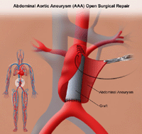
abdominal aortic aneurysm open repairA large incision is made in the abdomen to directly visualize the abdominal aorta and repair the aneurysm. A cylinder-like tube called a graft may be used to repair the aneurysm. Grafts are made of various materials such as Dacron (textile polyester synthetic graft) or polytetrafluoroethylene (PTFE, non-textile synthetic graft). This graft is sewn to the aorta, connecting one end of the aorta at the site of the aneurysm to the other end. The open repair is considered the surgical standard for an abdominal aortic aneurysm repair.
-
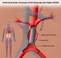
endovascular aneurysm repair (EVAR)EVAR is a procedure that requires only small incisions in the groinalong with the use of x-ray guidance and specially-designed instruments to repair the aneurysm. With the use of special endovascular instruments and x-ray images for guidance, a stent-graft is inserted via the femoral artery and advanced up into the aorta to the site of the aneurysm. A stent-graft is a long cylinder-like tube made of thin metal mesh framework (stent), while the graft is made of various materials such as Dacron or polytetrafluoroethylene (PTFE). The graft material may cover the stent. The stent helps to hold the graft open and in place.
 |
| Click Image to Enlarge |
Asymptomatic aneurysms may not require surgical intervention until they reach a certain size or are noted to be increasing in size over a certain period of time. Parameters considered when making surgical decisions include, but are not limited to, the following:
-
aneurysm size greater than 5 centimeters (about two inches)
-
aneurysm growth rate 0.5 centimeters (slightly less than one-fourth inch) over a period of six months to one year
-
patient’s ability to tolerate the procedure
For symptomatic aneurysms, immediate intervention is indicated.
Aortic Dissection
What is aortic dissection?
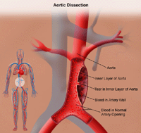
An aortic dissection begins with a tear in the inner layer of the aortic wall. The aortic wall is made up of three layers of tissue. When a tear occurs in the innermost layer of the aortic wall, blood is then channeled into the wall of the aorta, separating the layers of tissues. This generates great pressure in the aortic wall with a potential to rupture (burst). Aortic dissection can be a life-threatening emergency.
 |
| Click Image to Enlarge |
What causes aortic dissection?
The cause of aortic dissection is still under investigation. However, several risk factors associated with aortic dissection include, but are not limited to, the following:
-
hypertension (high blood pressure)
-
connective tissue disorders, such as Marfan’s disease, Ehlers-Danlos syndrome, and Turner’s syndrome
-
cystic medial disease (a degenerative disease of the aortic wall)
-
aortitis (inflammation of the aorta)
-
atherosclerosis
-
existing thoracic aneurysm
-
bicuspid aortic valve (presence of only two cusps, or leaflets, in the aortic valve, rather than the normal three cusps)
-
trauma
-
coarctation of the aorta (narrowing of the aorta)
-
hypervolemia (excess fluid or volume in the circulation)
-
polycystic kidney disease (a genetic disorder characterized by the growth of numerous cysts filled with fluid in the kidneys)
What are the symptoms of aortic dissection?
The most commonly reported symptom of an acute aortic dissection is severe, constant pain, sometimes described as “ripping” or “tearing,” and located in the chest, the middle of the abdomen, the lower back, or the pelvis area. The pain may be “migratory,” moving from one place to another, according to the direction and extent of the dissection.
The symptoms of aortic dissection may resemble other medical conditions or problems. Always consult your physician for more information.
How is aortic dissection diagnosed?
In addition to a complete medical history and physical examination, diagnostic procedures for an aortic dissection may include any, or a combination, of the following:
-
computed tomography scan (Also called a CT or CAT scan.) - a diagnostic imaging procedure that uses a combination of x-rays and computer technology to produce cross-sectional images (often called slices), both horizontally and vertically, of the body. A CT scan shows detailed images of any part of the body, including the bones, muscles, fat, and organs. CT scans are more detailed than general x-rays.
-
transesophageal echocardiogram (TEE) - a diagnostic procedure that uses echocardiography to assess the heart’s function and structures. A transesophageal echocardiogram is performed by inserting a probe with a transducer down the esophagus. By inserting the transducer in the esophagus, TEE provides a clearer image of the heart because the sound waves do not have to pass through skin, muscle, or bone tissue.
The physician will determine the most appropriate examination. When a diagnosis of aortic dissection is confirmed, immediate intervention is necessary. Medical intervention or surgery will be required depending on the location of the aortic dissection.
Click here to view theOnline Resources of Cardiovascular Disease